

Key takeaways: 3D CT imaging for facial surgical planning
- A 3D CT scan provides a rotatable, quantifiable digital model of the entire facial skeleton — not a surface approximation.
- It answers the decisive question that photo simulations cannot: is a concern caused by bone or by soft tissue?
- The data enables millimeter-precise planning of chin, cheek, and jaw augmentation or reduction, and custom implant design.
- It is indispensable for revision, asymmetry, and reconstructive cases, where the anatomy is compromised.
- Planning on skeletal truth reduces the risk of over-correction, under-correction, and misdiagnosis — the common causes of revision.
- It supports anatomy-based informed consent: patients see their own bone structure and understand exactly what is being planned.
The limitations of surface imaging: navigating beyond the illusion
Many technologies promise a preview of a future look by manipulating a photograph of the skin’s surface. While intriguing, these simulations share a critical flaw: they are entirely blind to the skeleton. They cannot reveal whether a weak chin is due to dental positioning or true mandibular retrognathia. They cannot measure the precise thickness of the malar bone to determine whether augmentation is needed or whether reduction is possible.
The most important variable in the plan is the one they cannot see. Planning with such tools is akin to an architect designing a building’s facade without knowing the load-bearing walls behind it — it risks a result that is cosmetically altered but structurally unsound or incongruent.
My philosophy rejects this superficial approach in favor of planning that begins with objective, deep anatomic data, a principle that is especially critical in revision and reconstructive facial surgery.
The 3D CT scan: a revelatory window to the skeletal truth
A 3D CT scan is a rapid diagnostic tool that transcends standard imaging, and where cone-beam CT is appropriate it produces the skeletal model at a considerably lower radiation dose than a conventional medical CT of the same region. It provides a volumetric, fully rotatable, and quantifiable digital model of the entire facial skeleton — not a surface approximation, but a precise mathematical representation of the underlying reality. Key metrics and revelations provided by this blueprint include:
- Bone prominence & volume: exact measurement of areas like the mandibular angles, the malar eminences, or frontal bossing. This answers the critical question: is the concern due to excess bone, or soft tissue?
- Bone deficiency & underdevelopment: objective identification of skeletal deficiencies in the chin (mentum), cheekbones, or jawline that no amount of soft-tissue filler can truly correct.
- Nasal architecture: quantification of septal deviation, dorsal hump composition (bone vs. cartilage), internal valve angles, and bone thickness for osteotomy planning.
- Asymmetry analysis: pinpointing the exact skeletal origin of facial asymmetry, distinguishing bone-based discrepancy from soft-tissue imbalance.
This data transforms the planning process from artistic estimation into a science of measurable goals.
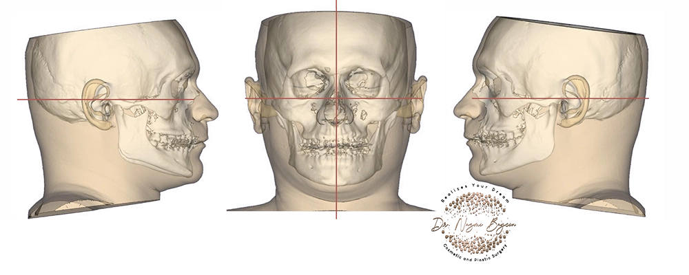
3D CT facial bone analysis — a quantifiable digital model of the facial skeleton used for precision surgical planning — by Dr. Nazmi Baycin, Dubai.
3D CT imaging versus surface photo simulation
| Capability | 3D CT imaging | Surface / photo simulation |
|---|---|---|
| Sees the bone | Yes — full skeletal model | No — skin surface only |
| Distinguishes bone vs. soft tissue | Yes, definitively | No |
| Millimeter measurement | Precise, quantifiable | Estimated / illustrative |
| Custom implant design | Enabled by exact topography | Not possible |
| Asymmetry & revision planning | Root-cause skeletal analysis | Superficial only |
| Best used for | Complex, structural, permanent alteration | General cosmetic previewing |
From digital data to surgical blueprint: the process of precision engineering
The scan is not an end, but a beginning. The subsequent analysis is where surgical strategy is born.
- Diagnostic clarity: I analyze the 3D model to diagnose the root cause. Is a “wide jaw” due to robust mandibular angles or masseter muscle hypertrophy? Is a “flat cheek” a result of thin soft tissue or a truly underprojected zygomatic bone? The scan provides the definitive answer.
- Virtual modeling & planning: using advanced software, I perform precise cephalometric analysis, virtually augmenting the chin or cheekbones by a specific number of millimeters and visualizing the new skeletal proportions in relation to the entire face. Conversely, I can simulate the reduction of a prominent mandibular angle or malar bone, calculating the exact volume of bone to be removed.
- Graft & implant design: the scan supplies the exact bony topography an implant has to sit against, which is what makes a patient-specific implant possible at all. What happens next — how that model becomes a printed implant — is a subject in its own right, and I set out the design-and-manufacturing stage in my article on custom 3D-printed facial implants in Dubai. Where a stock implant is the better choice instead, as in many cases of chin augmentation in Dubai, the same data guides selection and pocket planning.
This process ensures that the surgical plan addresses the true anatomic deficit or excess, not just its superficial manifestation.
Application in masterful practice: skeletal sculpting for harmony
The applications of this technology are transformative for specific surgical goals.
- Facial contouring & balancing: for chin augmentation (genioplasty) or reduction, determining whether an implant or a sliding osseous genioplasty is optimal and precisely calculating projection.
- Malar refinement: for cheekbone augmentation or reduction, differentiating soft-tissue midface deflation from true skeletal deficiency, so the correction is directed at the right layer.
- Upper facial third contouring: for a forehead that is flat or slopes backwards, an uneven brow ridge, or hollow temples, the scan measures frontal projection, brow ridge prominence, and temporal depth directly, so the correction is designed rather than estimated. How those measurements become a patient-specific frontal implant is the subject of my article on forehead bone augmentation in Dubai.
- Jawline sculpting: for mandibular angle reduction or a stronger, more defined jawline, precisely mapping the mandible to sculpt a smoother or stronger contour while preserving nerve integrity. How chin projection and jawline proportion are decided together is the subject of my article on chin and jawline structural harmony in Dubai.
- Complex rhinoplasty: indispensable for planning revision and major primary cases involving the bony pyramid.
- Post-traumatic & reconstructive surgery: rebuilding orbital walls, zygomatic arches, or the mandible based on mirror-images of the unaffected side or pre-injury scans.
The common alternative — operating without this skeletal map — risks over-correction, under-correction, or misdiagnosis, leading to patient dissatisfaction and the need for revision. The formal evidence for this comes from orthognathic surgery rather than from cosmetic facial contouring, and it is worth saying so plainly: what has been measured is jaw repositioning, and I am extending an established principle rather than citing a trial of my own procedures. Within that field the picture is consistent. A 2024 systematic review and meta-analysis comparing virtual surgical planning with conventional two-dimensional planning found that planning in three dimensions consistently narrows the gap between the planned bone position and the one actually achieved, particularly where the plan is carried into theatre with custom guides. The same analysis is candid about what it did not find: virtual planning showed a potential time advantage, but the difference in operating time was not statistically significant across the included studies. A companion review of 36 papers reaches the same conclusion on accuracy, describing precise translation of the plan and improved functional and aesthetic results. Accuracy is what the evidence supports here, not speed.
The ethical imperative: grounding consent in objective truth
Utilizing 3D CT imaging is, at its core, an ethical practice. It allows me to educate patients with undeniable visual evidence — I can show them where the chin is underprojected, or which side of the mandible is driving the imbalance they see in the mirror. This fosters informed consent based on anatomy, not abstract desire, and builds a partnership in which expectations are aligned with the precise surgical possibilities defined by the patient’s own bone structure. That transparency is fundamental to the trust I build with every patient in Dubai.
The standard of care for definitive certainty
The use of 3D CT imaging for surgical planning is reserved for complex facial skeletal surgery, where precision is decisive. It is employed when the diagnosis is unclear, the anatomy is compromised, or the procedure involves permanent alteration of the facial framework. The value lies in eliminating uncertainty, minimizing intraoperative surprises, and providing a clear, achievable roadmap for a result that is structurally sound, balanced, and intended to last a lifetime.
My philosophy is simple: beauty built on a foundation of truth is beauty that endures. The 3D CT scan is the ultimate expression of this principle — a tool that allows me to see, measure, and plan with the confidence of an architect who knows every detail of the ground upon which they build. For the discerning patient whose goals involve the fundamental reshaping of their facial architecture, this level of meticulous, evidence-based planning is not an optional extra; it is the standard of care.
FAQs about 3D CT imaging for facial surgery in Dubai
-
What is a 3D CT scan for facial surgery, and how is it different from a photo simulation?
A 3D CT scan creates a volumetric, rotatable, measurable digital model of your entire facial skeleton — the actual bone beneath the skin. A photo simulation only manipulates an image of the skin’s surface and is completely blind to the bone underneath. The difference is decisive: only a CT scan can tell whether a weak chin, flat cheek, or asymmetry comes from the bone or from the soft tissue, which determines the correct surgical plan.
-
Is a 3D CT scan safe? How much radiation is involved?
Cone-beam CT, which is the usual choice for facial skeletal planning, delivers a considerably lower radiation dose than a conventional medical CT of the same region, and the scan itself takes only moments. A conventional facial CT carries a higher dose, so it is reserved for cases where the additional detail genuinely changes the plan. In either case the dose is kept as low as is reasonably achievable while still producing an accurate skeletal model. For complex or permanent skeletal surgery, the diagnostic certainty is itself a safety benefit — it reduces the risk of misdiagnosis and of a second, revision operation.
-
Do I always need a 3D CT scan before facial surgery?
No. It is reserved for cases where it genuinely changes the plan — complex facial contouring, chin, cheek, or jaw skeletal work, revision surgery, significant asymmetry, major or revision rhinoplasty involving the bony pyramid, and reconstruction. Straightforward soft-tissue procedures usually do not require it. I recommend it only when the diagnosis is unclear, the anatomy is compromised, or the procedure permanently alters the facial framework.
-
Why can’t fillers or a photo preview replace this?
Fillers and surface previews address only the soft-tissue surface. If the underlying issue is skeletal — an underprojected chin, a deficient cheekbone, or an asymmetric jaw — no amount of filler truly corrects it, and a surface simulation cannot even reveal it. The 3D CT scan identifies the true anatomic cause so the correction is directed at the right layer rather than masking the surface.
-
Which procedures benefit most from 3D CT planning?
Chin augmentation or reduction (genioplasty), cheekbone (malar) augmentation or reduction, mandibular angle reduction and jawline enhancement, forehead and brow ridge contouring, complex and revision rhinoplasty involving the bony pyramid, correction of facial asymmetry, and post-traumatic or reconstructive surgery all benefit significantly. These are the cases where millimeter-level skeletal precision directly determines the quality and durability of the result.
-
How does 3D CT imaging help with facial asymmetry?
Facial asymmetry can be skeletal, soft-tissue, or both, and treating the wrong layer produces disappointing results. A 3D CT scan pinpoints the exact skeletal origin of the asymmetry and allows the unaffected side to be mirror-imaged as a template for correcting the affected side. This turns asymmetry correction from an estimate into a measured, root-cause plan.
-
How does the scan help design a custom facial implant?
Because the CT data captures your unique skeletal topography exactly, a custom implant can be designed or selected to fit your bone precisely, rather than adapting a generic off-the-shelf implant during surgery. A better-fitting implant integrates more reliably, sits more stably, and produces a more natural, predictable contour.
-
Does 3D CT planning make surgery safer and more predictable?
Yes. Planning on an accurate skeletal model reduces intraoperative surprises, helps preserve important structures such as nerves, and lowers the risk of over-correction, under-correction, or misdiagnosis — the usual reasons a revision becomes necessary. The published evidence on 3D virtual planning comes mainly from orthognathic surgery, where it reports better accuracy in translating the plan onto the bone and improved functional and aesthetic outcomes. It does not demonstrate a significant saving in operating time, and I would not claim one.
The architecture of confidence
For the discerning patient in Dubai whose goals involve reshaping their facial architecture, this evidence-based planning is the assurance that the changes we discuss are not just wishes, but carefully engineered plans derived from the reality of their unique anatomy. To explore how this commitment to precision can guide your transformative journey, I invite you to learn more about my approach as a leading facial aesthetic and reconstructive surgeon in Dubai.


GET APPOINTMENT
Get ready to look and feel best… You deserve…
