

Key takeaways: the ear is three subunits, not one shape
- Prominence is an anatomical diagnosis, not a single condition with a single cure.
- The antihelix is the inner fold that holds the outer rim back; when it is weak, the rim has nothing to fold against.
- The concha is the deep central bowl; when it is over-deep it acts as a spacer, pushing the whole ear out.
- The lobule contains no cartilage at all, which is why it behaves differently from the rest of the ear.
- The ideal ear sits roughly 15–20 mm from the head with a natural antihelical fold.
- Most prominent ears combine two or three subunit problems, in different proportions on each side.
This article is about the anatomy — what each subunit is and what goes wrong with it. How that anatomy is corrected, step by step in the operating room, is set out separately in my article on the surgical sequence of otoplasty. For the procedure as a whole — candidacy, anesthesia, recovery and cost — see my main page on otoplasty in Dubai.
Reading the ear: one silhouette, three structures
The ear’s prominence is judged not in isolation, but in relation to the head. The ideal ear sits approximately 15–20 mm from the side of the head at the midpoint, with a gentle, parallel alignment to the profile. When that distance is exceeded, or when specific contours are absent, the ear is perceived as protruding or cup-shaped. What matters clinically is that the same visible result can arise from entirely different causes.
The StatPearls reference chapter on otoplasty puts the anatomy plainly: prominauris, the most common reason for ear correction, is generally caused by excessive conchal bowl depth and effacement of the antihelix. Two structures, either or both. Add the lobule, which follows its own rules, and the diagnostic picture is complete.
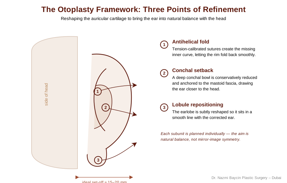
The otoplasty framework: creating the antihelical fold, setting back the conchal bowl, and repositioning the lobule for natural balance — by Dr. Nazmi Baycin, Dubai.
The antihelix: the fold that holds the rim back
The antihelix is the curvilinear inner ridge that separates the outer rim, or helix, from the conchal bowl. Superiorly it divides into a superior and an inferior crus, which together with the helix define the triangular fossa; inferiorly it ends at the antitragus. Its job is structural. A well-formed antihelix is what allows the helical rim to fold gracefully back toward the head.
When the fold is weak or effaced, the rim has nothing to fold against and stands away on its own. This is why an ear can protrude even when the conchal bowl is of perfectly normal depth. It is also why the inferior crus is usually already well formed even in severe prominence, while the superior crus is the part that is commonly missing — a detail that matters when planning where a new fold needs to sit.
The concha: the bowl that acts as a spacer
The concha is the deep, cup-shaped cavity beside the ear canal, divided by the crus of the helix into the cymba concha above and the cavum concha below. Unlike the antihelix, it does not shape the rim. It sets the distance.
An excessively deep concha works as a structural spacer between the ear and the skull, pushing the entire auricle outward regardless of how good the antihelical fold is. An ear with conchal excess cannot be brought into balance by addressing the fold alone; the depth itself is what has to change. In practice the conchomastoid angle is approximately 90 degrees in a normal ear, and it is the widening of that relationship, rather than the ear’s own shape, that drives this pattern of prominence.
The lobule: the subunit with no cartilage in it
The lobule is the exception in every sense. It is the one part of the auricle that contains no cartilage at all, and it does not develop from the same embryological structures as the rest of the ear. That single anatomical fact explains most of what goes wrong with it.
Because it holds no cartilage, the lobule cannot be reshaped by any of the maneuvers that work on the antihelix or the concha. It has no spring to release and no fold to create. Its position is instead governed by what sits above it — chiefly the cauda helicis, the cartilaginous tail of the helix, which can continue to press the lobe outward even after the upper two-thirds of the ear have been brought perfectly into place. StatPearls records this as a recognized late problem after otoplasty, the outstanding lobule, and notes that removing the cauda helicis is what usually resolves it.
| Subunit | What it is | What goes wrong |
|---|---|---|
| Antihelix (inner fold) | The curved inner ridge dividing the rim from the bowl, dividing above into superior and inferior crura | Weak or effaced fold, most often the superior crus, so the rim has nothing to fold against and stands out on its own |
| Concha (central bowl) | The deep cavity beside the ear canal, sitting at roughly a 90-degree angle to the mastoid | Excess depth acts as a spacer, pushing the whole ear outward however good the fold above it is |
| Lobule (earlobe) | The soft lower third, containing no cartilage and developing separately from the rest of the auricle | Held outward by the cauda helicis above it, so it can stay prominent even after the cartilaginous ear is corrected |
Why the lobule is the subunit most often missed
Of the three, the lobule is the one most often left out of the plan, and the reason is structural rather than careless. The antihelix and the concha are cartilage problems, and cartilage is what an otoplasty is built around. The lobule sits outside that logic entirely.
The result is a specific and recognizable outcome: an ear whose upper two-thirds sit neatly against the head while the lobe still stands away, giving a bottom-heavy silhouette that reads as unfinished from the front. Occasionally the medialization of the auricle even has a paradoxical effect, drawing the upper ear back while the lobe swings further out. Because the lobe cannot be folded, the correction has to come from the cartilage above it and from careful reshaping of the soft tissue — which is why it is planned as a subunit in its own right, from the outset, rather than assessed at the end.
Anatomical variation: why your two ears are not the same problem
Prominence is rarely symmetrical in its cause. One ear may protrude because the antihelix never formed properly, while the other protrudes because the conchal bowl is unusually deep, and the two will look broadly similar from the front while requiring different answers.
This is the practical value of thinking in subunits. Assessing each ear against the same three questions — is the fold present, is the bowl too deep, is the lobe held out — produces a diagnosis for each side independently. It also sets a realistic goal. Perfect symmetry does not exist in unoperated ears; what is achievable is dynamic symmetry, where both ears read as a balanced pair even though their underlying anatomy differed. Cartilage stiffness also varies with age, which changes how each of these structures behaves under the knife; I cover that in my article on how cartilage behaves differently in children and adults.
Diagnosis before technique
Otoplasty is a quiet procedure with a loud impact. It operates on the periphery of the face, yet its effect on a person’s frontal confidence is central. What decides that outcome is not which instrument is chosen but whether the ear was correctly read in the first place — because a beautifully executed antihelical fold does nothing for an ear whose prominence was coming from the bowl, and a perfectly set-back concha leaves a bottom-heavy result if the lobe was never part of the plan. Anatomy first, then technique. To discuss which subunits are driving prominence in your own ears, or your child’s, I welcome you to consult with me as a leading cosmetic surgeon in Dubai.
FAQs about auricular anatomy and prominent ears
-
Which parts of the ear actually cause it to stick out?
Three subunits, working alone or together. The antihelix is the inner fold that lets the outer rim curl back toward the head, and when it is weak the rim stands away on its own. The concha is the deep bowl beside the ear canal, and when it is too deep it acts as a spacer that pushes the whole ear outward. The lobule is the soft lower third, which can remain prominent even after the cartilage above it is corrected. Most prominent ears involve at least two of the three, in different proportions on each side.
-
What does the antihelical fold actually do?
It is the structural element that holds the outer rim back. The antihelix is the curved inner ridge between the rim and the bowl, dividing above into two crura that frame the triangular fossa. A well-formed one gives the rim something to fold against, which is what produces the ear’s natural three-dimensional depth. When it is effaced, the rim has no support and protrudes independently of everything else. In severe prominence the lower crus is usually still present and it is the upper one that is missing, which is why the top of the ear is so often the part that stands out most.
-
How can an ear stick out if the fold looks normal?
Because the fold and the distance are governed by different structures. The antihelix shapes the rim; the concha sets how far the whole ear sits from the skull. An unusually deep conchal bowl works as a spacer between ear and head, so the auricle is pushed outward even though its own contours are perfectly well formed. Ears in this pattern often look correctly shaped in profile and clearly prominent from the front. Recognizing it matters because no amount of work on the fold will bring the ear closer if the depth of the bowl is what is holding it out.
-
Why does the earlobe behave differently from the rest of the ear?
Because it contains no cartilage. The lobule is the only part of the auricle without a cartilaginous framework, and it develops separately from the rest of the ear in the embryo. That means it has no spring to release and no fold that can be created in it. Its position is dictated instead by the cartilage above it, particularly the tail of the helix, which can keep pressing it outward after the upper ear has been brought into place. So the lobe is never corrected by reshaping it directly, which is exactly why it needs to be identified as its own subunit at the assessment stage.
-
Can my two ears need different corrections?
Very commonly, yes. Two ears can protrude by a similar amount for entirely different anatomical reasons — one because the antihelical fold never formed, the other because the conchal bowl is deep. From the front they look like the same problem; structurally they are not. Each ear is therefore assessed on its own against the same three questions, and the plan can legitimately differ between sides. The goal is balance between the pair rather than two identical structures, because unoperated ears are never perfectly symmetrical either.
-
How far from the head should a normal ear sit?
As a working reference, roughly 15 to 20 millimetres at the midpoint, with the ear aligned gently parallel to the profile. It is worth treating that as a guide rather than a target, for two reasons. Published normal ranges vary depending on whether the measurement is taken at the upper pole, the mid rim, or the lobe, since the ear is not equidistant from the skull along its length. And prominence is often defined by the angle between the ear and the head rather than by a distance at all. What matters more than any single number is that the ear looks proportionate to the head it belongs to.


GET APPOINTMENT
Get ready to look and feel best… You deserve…
