

Key takeaways: rebuilding, not revising
- Secondary rhinoplasty is reconstruction in a scarred field, not a simple touch-up.
- The rebuild uses autologous (your own) cartilage, which integrates as living tissue.
- Grafts follow a hierarchy: septal, then auricular (ear), then costal (rib) cartilage.
- Every graft is dual-purpose — a spreader graft is both an aesthetic spacer and an airway stent.
- Synthetic implants and allografts resist integration and risk extrusion in a revision field.
- Healing is slower; the final contour can take 18–24 months, and staging is sometimes safest.
The anatomical legacy of primary surgery: diagnosing the deficit
A systematic analysis is the cornerstone of any successful secondary rhinoplasty in Dubai. The deficiencies I encounter form a predictable catalog of structural failure. The over-resected dorsum — excessive removal of dorsal septal cartilage — creates a low, scooped saddle nose and often destabilizes the middle third, causing internal valve collapse and difficulty breathing. In revision cases I see in Dubai, the weakened tip, from aggressive removal of the lower lateral cartilages or inadequate primary support, results in a pinched, under-projected, or drooping tip.
The collapsed mid-vault, from poor preservation of the upper lateral cartilages, narrows the middle third and is the primary cause of persistent nasal airway obstruction after rhinoplasty, as the internal valves collapse inward on inspiration. And the scarred lining — internal synechiae or compromised mucosa — restricts skin drape and limits reconstructive options. Each issue requires not camouflage but structural reinforcement, which is exactly where synthetic materials and allografts consistently fail: they cannot integrate, resist infection poorly, and often lead to extrusion or visible, unnatural contours.
Why these deficits arise, how the choice of donor material is reasoned through, and why irradiated and alloplastic materials are avoided in a scarred bed are set out in my article on advanced techniques for revision rhinoplasty. What follows here is narrower: the graft-by-graft sequence by which the framework is actually rebuilt.
The autologous armamentarium: choosing the right graft material
The philosophy of using the patient’s own tissue rests on biological integration: these grafts become vascularized, living parts of the new framework. My selection follows a hierarchy based on need and availability, summarized below. Where the graft is shaped from fragmented cartilage, technique matters for a smooth, stable contour; a 2024 systematic review of thirty studies of mechanically fragmented cartilage grafts examines how diced, shaved and crushed cartilage behave as grafting material. The principle of combining structural and diced cartilage for the dorsum is one I have long worked with in my own practice, described in the 2004 paper I co-authored on a bone-and-diced-cartilage graft for the nasal dorsum.
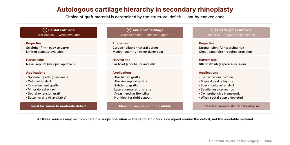
The autologous reconstruction of the nasal framework in secondary rhinoplasty — graft sources (septal, ear, and rib cartilage) and their placement as dorsal onlay, spreader, tip-support, and alar batten grafts — by Dr. Nazmi Baycin, Dubai.
Septal cartilage is the ideal source when enough remains — straight, strong, and easy to shape. Auricular cartilage is softer and pliable, perfect for alar rim repair or subtle tip work. Costal (rib) cartilage is the powerhouse of revision surgery: when significant structure is needed, I harvest a segment of the patient’s own rib and sculpt it precisely, minimizing warping through careful carving. Harvest is integrated into a holistic plan for composite nose surgery in Dubai, with donor sites closed to minimize visibility.
| Graft source | Character | Best used for |
|---|---|---|
| Septal cartilage | Straight, strong, easy to shape | First choice: spreader grafts, strapping grafts, minor tip refinements |
| Auricular (ear) cartilage | Softer, pliable, naturally curved | Alar rim defects, subtle tip grafts, areas needing flexibility |
| Costal (rib) cartilage | Powerful, abundant, strong | Major support: rebuilding a collapsed dorsum, new L-strut, major tip grafts |
The reconstructive sequence: engineering a new foundation
The surgery follows a logical, stepwise sequence, rebuilding the nose from the inside out.
Meticulous exposure and analysis
The approach is almost always an open rhinoplasty, giving the complete visualization needed to assess the damage. Scar tissue is carefully released, and the remaining anatomical remnants are meticulously preserved.
Re-establishing the dorsal and mid-vault lines
For the collapsed dorsum, I fashion a strong dorsal onlay graft from rib cartilage, carved to a natural, slightly tapered profile. To open the collapsed internal valves, I insert bilateral spreader grafts — long, thin strips of cartilage between the septum and upper lateral cartilages. This simultaneously widens the pinched middle third and restores the physiologic airflow triangle of the internal valve.
Rebuilding the tip complex
A weak, unsupported tip requires a foundational strategy. I frequently use a septal extension graft or a columellar strut of strong rib or septal cartilage, fixed securely to the caudal septum as a new pillar. To this stable base I suture precisely carved tip grafts to define projection, rotation, and contour, ensuring a tip that is resilient and will not droop over time.
Reinforcing the alar rims and external valves
Collapsed or retracted nostrils need reinforcement. I use thin, curved strips of ear cartilage as alar batten grafts, placed in precise pockets along the alar rim to provide support without stiffness — restoring both the external contour and the function of the external nasal valve.
The synergy of form and function: the biomechanics of a living framework
The ultimate goal is a nose that breathes as well as it looks. Every graft serves a dual purpose. A spreader graft is both an aesthetic spacer and a functional stent for the airway. A strong columellar strut provides tip projection and prevents dynamic collapse on deep inspiration. By rebuilding with integrated autologous tissue, I create a framework that responds to facial animation, trauma, and aging as a unified, living structure. Why that inseparability of form and function matters to the patient, and how the assessment and the recovery timeline follow from it, I set out in my article on restoring form and function in secondary rhinoplasty. Where residual septal deviation contributes to obstruction, it is corrected concurrently; to understand that functional component, visit septoplasty surgery in Dubai.
The realism of revision: managing expectations and staging
Secondary rhinoplasty requires profound surgical patience — during the operation and in managing expectations. Healing is longer and less predictable because of pre-existing scar tissue, and I counsel patients that the final contour may take 18–24 months to fully refine. In cases of severe damage or compromised skin, a staged approach may be safest: the first stage restores the structural foundation and internal function, and a later, minor stage addresses final refinements of contour.
Honesty about this process is a critical component of the trust such a complex journey requires. Because each reconstruction is individual, its plan and cost are set out transparently at consultation. This restorative philosophy runs through all of my facial plastic surgery work in Dubai.
The art of restoration
Secondary rhinoplasty is the ultimate test of a surgeon’s skill, and in my secondary rhinoplasty practice in Dubai it moves the surgeon from the role of sculptor to that of architect and engineer. It demands a deep reverence for nasal physiology, mastery of graft harvest and fabrication, and the artistic vision to see harmony within a scarred landscape. My commitment is to leverage the body’s own biological wisdom — using autologous tissue — to build a nose that is not only beautiful and proportionate, but strong, functional, and permanently integrated. It is a restorative procedure that heals more than the nose: it restores confidence and the simple, vital ability to breathe freely.
FAQs about secondary rhinoplasty in Dubai
-
How do I know if I need secondary rhinoplasty, or if I should wait longer after my primary?
The minimum waiting period before revision is twelve months from the primary surgery, and in most cases eighteen months is more informative. The nose continues to change — swelling resolves in stages, scars contract, and cartilage grafts settle — over this entire period. What appears to be a persistent deformity at six months may partially or fully resolve by month fifteen. I advise patients to document their nose photographically every three months and to come for reassessment when the appearance has been stable for at least three to four consecutive months, as this indicates the final result has been reached.
-
What is a spreader graft, and why is it so important in revision surgery?
A spreader graft is a thin, rectangular strip of cartilage placed between the nasal septum and the upper lateral cartilage on each side. It serves two purposes simultaneously: aesthetically it widens a pinched middle third, restoring the smooth dorsal lines; functionally it opens the internal nasal valve — the critical airflow triangle between the septum and the upper lateral cartilage — which is the most common site of airway obstruction after over-aggressive primary rhinoplasty. A spreader graft is both a structural correction and a functional restoration in a single maneuver.
-
Why is rib cartilage used, and is a second operation site necessary?
Rib cartilage is needed when the structural deficit exceeds what septal or ear cartilage can supply — typically in cases of saddle nose deformity, collapsed mid-vault requiring major bilateral spreader grafts, or a tip with no remaining cartilaginous support. Yes, it requires a separate incision on the chest, typically 2-3cm, placed in or below the inframammary fold where it is well-concealed. This additional scar is a trade-off that virtually all patients readily accept when the alternative is an inadequate reconstruction with insufficient material.
-
How is each graft shaped and fixed so it does not move or warp?
Warping is the principal technical risk with rib cartilage, and it is managed at the carving stage rather than afterwards. I carve concentrically from the balanced central portion of the segment, allow the carved graft to sit before final shaping so that any tendency to bend declares itself, and fix the graft securely rather than relying on a press fit. A dorsal onlay is seated on a precisely dissected bed so it cannot slide; a columellar strut or septal extension graft is fixed to the caudal septum so the tip has a fixed pillar rather than a floating one; and alar battens sit in pockets cut to their exact dimensions. Fragmented cartilage behaves differently again, which is why the technique used to dice or crush it matters for the smoothness of the final contour.
-
Why does secondary rhinoplasty take longer to heal than a primary procedure?
Because the tissue environment is fundamentally different. Scar tissue from the primary surgery creates a less compliant, less vascular environment that is slower to reduce swelling and less predictable in its healing behavior. New grafts must integrate into an already-altered scaffold. The skin envelope has lost some elasticity and may take longer to redrape over the new framework. For all these reasons, the eighteen to twenty-four month timeline to final result that I counsel for secondary cases is not pessimism — it is an honest and accurate description of the biology of scar tissue healing.
-
What is the risk that revision surgery will make things worse?
It is a real risk that I discuss explicitly with every revision patient. Operating in a scarred field introduces unpredictability — scar tissue can bleed unexpectedly, planes that should be easily separated are adherent, and the skin may not redrape exactly as planned. This is why I strongly prefer to wait for full maturation before operating, stage complex cases where necessary, and operate with a conservative philosophy: it is always better to achieve a meaningful, reliable improvement than to pursue perfection and risk destabilizing a stable result. The goal of revision is improvement, not perfection in a single operation.


GET APPOINTMENT
Get ready to look and feel best… You deserve…
