

Key takeaways: orbicularis suspension in lower blepharoplasty
- The lower eyelid is a dynamic structure to be supported, not simply a flap of skin to be trimmed.
- With age the orbicularis oculi muscle descends, dragging the lid down and creating laxity, festoons, and a deepened lid-cheek junction.
- Suspending the muscle — hitching it to a stable higher point — recreates the internal sling that youth provides naturally.
- The orbicularis hitch achieves this with a single suture; a flap suspension gives more robust support in older patients.
- Restoring muscle support lets skin be excised conservatively, reducing the risk of lid retraction and scleral show.
- The philosophy mirrors modern facelifting: preserve and support, not remove and tension.
The anatomical rationale: why suspension matters
To appreciate why orbicularis suspension is so effective, one must understand the anatomy of the aging lower lid. The orbicularis oculi muscle is divided into two functional parts: the pretarsal portion, which overlies the tarsal plate and drives rapid blinking, and the preseptal portion, which lies over the orbital septum and provides voluntary lid closure and support.
With age, the preseptal orbicularis descends, and this descent contributes directly to lid laxity, festoons (malar mounds), and the deepening of the lid-cheek junction. Once the muscle descends, it no longer supports the overlying skin and underlying septum — producing a tired, aged appearance that simple skin excision cannot correct.
Orbicularis suspension addresses this problem at its source. By elevating the descended muscle and securing it to the periosteum of the lateral orbital rim or the deep temporal fascia, the surgeon recreates the supportive sling that youth provides naturally. The elevated muscle lifts the overlying skin, smooths the lid-cheek junction, and provides durable support that prevents recurrent laxity.
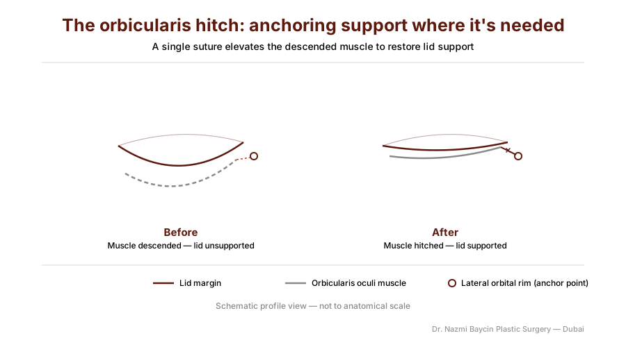
The orbicularis hitch mechanism: a single suture elevates the descended preseptal muscle to the lateral orbital rim, forming an internal sling that supports the lower lid — by Dr. Nazmi Baycin, Dubai.
The suspension techniques
The concept of suspending the orbicularis is not new, but the techniques have evolved significantly. The earliest descriptions involved extensive dissection and formal myotomies — cutting through the muscle to create a flap that could be repositioned. While effective, these approaches carried risks of bleeding, denervation, and prolonged swelling. An orbicularis transposition flap technique described from Montevideo in 2006 provided vertical support through a laterally based muscle flap, elevating a strip of orbicularis and transposing it to the lateral orbital rim to create a muscular sling — a significant advance, but still requiring substantial dissection. The modern era has been defined by a movement toward simplicity: achieving the benefits of muscle support without the morbidity of extensive dissection.
That movement runs in two directions, and it is worth being open about the disagreement. One widely described modern variant pairs orbicularis suspension with lateral retinacular suspension through a transconjunctival approach, and its authors argue against the tradition this article works within: that extensive orbicularis dissection, and the commonly performed skin-muscle flap, denervate the muscle without improving the aesthetic result. They report that their technique improves the lid-cheek junction and the skin and maintains or improves the natural posture of the lower lid — though as a technique description rather than a series with outcome data. My own position is narrower than either camp: where the skin itself must be addressed, a skin-muscle flap earns its place; where it does not, the transconjunctival route is the lighter operation.
The orbicularis hitch: simplicity and efficacy
Among the most elegant modern techniques, the orbicularis hitch achieves muscle suspension through a single suture, without significant myotomy or lateral canthal manipulation. The technique is deceptively simple: after completing the standard skin-muscle flap dissection, the surgeon identifies the descended preseptal orbicularis, and a single, strategically placed suture gathers a small amount of this muscle and secures it to the periosteum of the lateral orbital rim, just inside the orbital tubercle. This single hitch elevates the entire muscle sheet, lifting the lid and restoring its youthful contour.
The published experience is encouraging, with the caveats that matter. A retrospective review of 100 lower blepharoplasties by orbicularis hitch (Little & Hartstein) was carried out as two consecutive series of 50, one by a plastic surgeon and one by an oculoplastic surgeon, with mean skin excision of 8 mm and 5 mm and average follow-up of 17 and 16 months. Outcomes were reported per lid across 200 lids: lateral canthal support was added to 2.5% of lids, and there was a single lid malposition (0.5%) requiring surgical revision, with no other complication or reoperation.
The authors concluded that the hitch provides the benefits of muscle suspension through a simple, single-suture elevation of descended muscle, while maintaining lid support and lateral canthal integrity. Two things belong alongside that conclusion. The journal grades the paper Level of Evidence 4 — a retrospective, single-arm series with no comparison group, assessed by the two operating surgeons themselves. And the conclusion drew a published rebuttal in the same journal, in which two oculoplastic surgeons argued that the data presented do not support it, noting that some patients nonetheless underwent tarsopexy, canthoplasty or canthopexy despite the claim that the hitch nearly eliminates the need for horizontal lid tightening. I find the technique valuable in my own hands; that is a clinical judgement the series supports rather than proves.
The orbicularis flap suspension: evidence in the older patient
For the older patient, in whom tissue descent is more pronounced and skin quality poorer, a more robust suspension may be indicated. A retrospective single-centre study of 39 patients aged 60 and over (average age 67.5 years) performed orbicularis oculi flap suspension alongside transcutaneous blepharoplasty to treat festoons, fat prolapse, and lid laxity. Eyelid laxity was corrected in every patient with no recurrence recorded, complications were minimal (transient skin edema in two patients, transient ectropion in one), and subjective satisfaction averaged 2.56 out of 3.
Two qualifications matter when reading those numbers. Follow-up averaged 145.7 days — under five months — so the absence of recurrence describes early stability rather than durability. And like the hitch series, the study has no comparison group, so it documents what the technique achieved in these hands rather than showing it to be better than an alternative.
The authors highlighted the technique’s surgical ease and reproducibility, a shortened operation time, the avoidance of conjunctival complications since no transconjunctival approach is used, and reliable correction of the underlying laxity.
Technical variations: matching suspension to anatomy
No single suspension technique is optimal for every patient; the choice must be guided by anatomy and the degree of lid laxity.
| Technique | Description | Best indications | Key evidence |
|---|---|---|---|
| Orbicularis hitch | Single-suture suspension of preseptal muscle to the lateral orbital rim | Mild to moderate lid laxity; primary blepharoplasty | Little & Hartstein: one lid malposition in 200 lids (0.5%) |
| Orbicularis flap suspension | More extensive mobilization and suspension of an orbicularis flap | Severe laxity; patients over 60; festoons | Lee et al.: laxity corrected in all 39 patients, mean follow-up 146 days |
| Orbicularis transposition flap | Laterally based muscle flap transposed to the orbital rim | Combined lid and midface descent | Carriquiry et al.: technique description, no comparative outcome data |
| Combined with canthopexy | Suspension plus canthal tightening | Significant horizontal laxity; negative-vector patients | Canthal support was added to 2.5% of lids in the Little series |
In my practice, I assess each patient’s lid laxity, skin quality, and orbital vector before selecting the technique. For most primary blepharoplasty patients, a modified orbicularis hitch provides excellent support with minimal invasiveness; for the older patient with significant descent or festoons, a more formal flap suspension may be indicated.
Patient selection: identifying the candidate for suspension
The findings that point specifically to muscle suspension are muscular ones: festoons or malar mounds, a lid-cheek junction that has visibly deepened, and an orbicularis that has descended out of position. Where those are present, elevating the muscle treats the cause rather than the surface, and no amount of skin excision will substitute for it.
The wider preoperative risk assessment sits alongside that and answers a different question. Vector analysis, the snap-back and distraction tests, midface descent and skin quality together decide how much support the lateral canthus needs, and I set that assessment out in full in preventing retraction through canthal support. Both are done at the same consultation: one asks what the muscle is doing, the other asks what the tendon can hold.
Combining suspension with other maneuvers
Orbicularis suspension does not exist in isolation; in a comprehensive lower blepharoplasty it is combined with other maneuvers. Rather than excising herniated fat, I prefer to preserve and reposition it to fill the tear-trough hollow — the same logic behind how the fat is repositioned to fill the tear-trough hollow — with the elevated orbicularis providing a supportive bed for the repositioned fat.
With muscle support restored, skin can be excised conservatively — the elevated muscle lifts the skin, reducing how much must be removed and minimizing the risk of lid retraction. And in patients with significant horizontal laxity, canthopexy or canthoplasty may be added to the suspension, which is the focus of how canthal support prevents lid retraction.
The philosophy of proactive support
My philosophy in lower blepharoplasty has evolved from remove and close to preserve and support. The orbicularis muscle is not merely tissue to be divided and discarded; it is a dynamic structure that, when properly supported, becomes the foundation of lasting lid rejuvenation.
This aligns with the broader trend in facial aesthetic surgery toward preservation and suspension rather than excision and tension — just as a deep-plane facelift preserves the SMAS and lifts the descended midface, orbicularis suspension preserves the muscle and lifts the descended lid. The result is not a pulled, operated appearance but a naturally refreshed, youthful contour. Patients interested in how these techniques apply to their own anatomy can learn more about my comprehensive approach to eyelid surgery in Dubai.
FAQs about orbicularis suspension lower blepharoplasty in Dubai
-
What is orbicularis suspension, and how is it different from standard lower blepharoplasty?
Standard lower blepharoplasty removes excess skin and fat and closes the incision, which can work well in younger lids with good tone. Orbicularis suspension goes a step further by lifting the descended orbicularis muscle and anchoring it to a stable point on the orbital rim, recreating the natural internal sling that supports the lid. This treats the underlying cause of lid descent rather than just the surface signs, giving a more natural and longer-lasting result.
-
Does supporting the muscle change how much skin has to be removed?
Yes, and that is much of the point. With the orbicularis elevated and anchored, the muscle carries the lift, so less skin needs to come out and the closure is not left under vertical tension. Skin removal becomes the last step and the smallest one. How the lateral canthus is assessed and supported alongside this, and when a canthopexy or canthoplasty is indicated, is set out separately in my article on preventing retraction through canthal support.
-
How do you decide whether a lid needs muscle suspension?
By how the muscle itself is behaving. Visible festoons or malar mounds, a lid-cheek junction that has deepened, and an orbicularis that has clearly descended all point toward suspension, and I assess them at consultation. The wider preoperative risk assessment that decides the canthal side of the plan — vector analysis, the snap-back and distraction tests, midface descent and skin quality — is set out in my article on preventing retraction through canthal support, since those findings govern both halves of the operation.
-
Is the orbicularis hitch or the flap suspension better?
Neither is universally better — they suit different anatomy. The orbicularis hitch is a single-suture technique ideal for mild to moderate laxity and primary surgery, offering strong support with minimal invasiveness. A flap suspension is more robust and better suited to older patients with pronounced descent, poorer skin quality, or festoons. Part of the consultation is matching the right level of support to your specific lid.
-
Will my eye shape or gaze look changed or operated on?
The goal is the opposite of an operated look. Because the technique restores the lid’s own support rather than pulling skin tight, it aims to preserve your natural eye shape and gaze while refreshing the area. The lid is elevated to a youthful position rather than stretched, so the result reads as rested and natural rather than tight or altered.
-
How long is recovery after orbicularis suspension blepharoplasty?
Most patients have moderate swelling and bruising for the first week and return to work and social activities within about 10 to 14 days. A temporary feeling of tightness at the outer corner of the eye is normal and settles as the tissues soften. The final contour continues to refine over several months as swelling fully resolves and the lifted lid-cheek junction becomes apparent.
-
Can suspension be combined with fat repositioning or a canthal procedure?
Yes, and often it should be. Rather than removing herniated fat, it can be preserved and repositioned to fill a tear-trough hollow, with the lifted muscle providing a supportive bed. In patients with significant horizontal laxity, a canthopexy or canthoplasty may be added to tighten the lid further. These maneuvers are planned together so the whole lower lid and lid-cheek junction are addressed as one unit.
-
Is orbicularis suspension safe?
In experienced hands it has a strong safety record. Published series report low complication rates — for the single-suture hitch, a lid malposition rate of around 0.5%, and for flap suspension, only transient, self-resolving issues such as short-lived swelling. As with any eyelid surgery, results and safety depend heavily on proper patient selection and surgical experience, which is why careful preoperative assessment matters so much.
Choose support, choose natural results
The lower eyelid is a dynamic structure, not a static flap — it blinks, it squints, it expresses emotion, and it deserves a surgical approach that respects its function while enhancing its form. I have made orbicularis suspension a cornerstone of my lower blepharoplasty technique because the published experience supports it: whether through the elegant simplicity of the orbicularis hitch or the robust support of the orbicularis flap, muscle suspension delivers well-supported lids with low reported complication rates. That evidence describes what the technique achieves rather than proving it superior to every alternative, and my preference for it rests on those series together with my own results. If you are considering lower blepharoplasty and seek a surgeon who will support your lid rather than simply trim it, I invite you to explore my philosophy of plastic surgery in Dubai and schedule a consultation.


GET APPOINTMENT
Get ready to look and feel best… You deserve…
