

Key takeaways: pedicle choice and nipple sensation
- The nipple-areola complex is a neurosensory organ, so sensation deserves a deliberate preservation strategy.
- Sensation is carried mainly by the 4th intercostal nerve (with 3rd and 5th), running superomedial to inferolateral.
- The pedicle is a protected neural corridor, not just a blood supply — its choice is the main variable under the surgeon’s control.
- The superomedial pedicle aligns with the dominant nerve’s course; the evidence on sensation is favorable but genuinely mixed.
- The free nipple graft severs all neural connections and is reserved for extreme cases only.
- Nerve recovery is nonlinear, with the final baseline typically set by 18–24 months.
The neural cartography of the breast
Preservation begins with precise anatomical understanding. The primary sensory innervation of the nipple-areola complex (NAC) derives from the lateral and anterior cutaneous branches of the third, fourth, and fifth intercostal nerves, with the fourth typically dominant. Crucially, these nerves do not travel in isolation; they course within the parenchyma alongside the vascular supply, often in a superomedial-to-inferolateral orientation.
The surgical error that leads to sensory loss is typically one of disruption, stretch, or devascularization of these fine neural filaments — a pedicle that is too long, undergoes excessive torsion, or is dissected in a plane that severs these pathways will compromise sensation. My preoperative planning therefore visualizes this corridor in three dimensions, determining which approach best aligns with and safeguards each patient’s inherent neural anatomy.
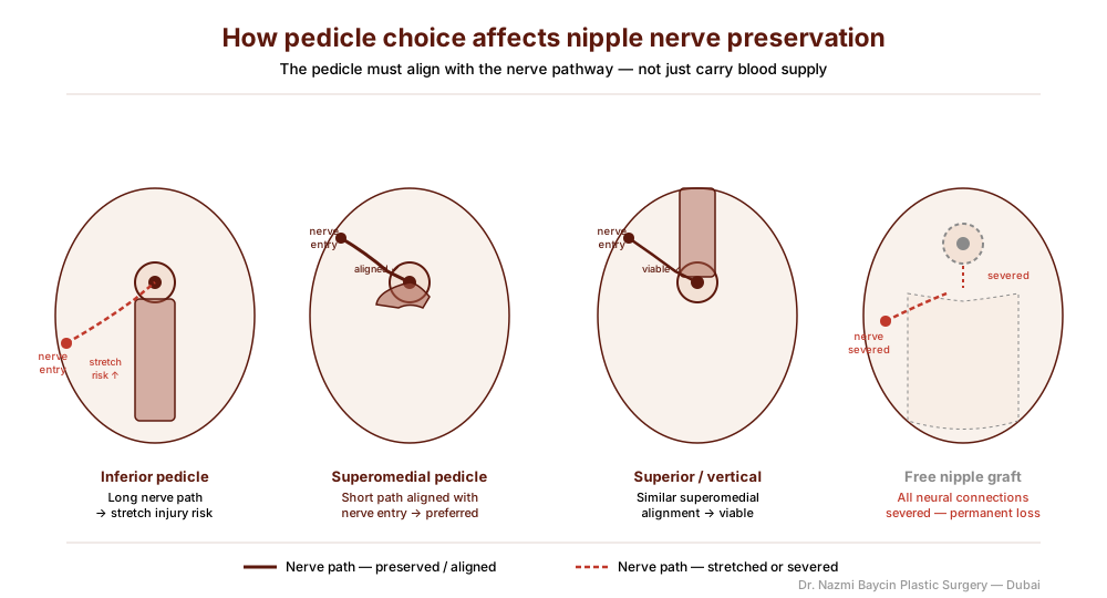
A comparison of the main pedicle techniques and how each relates to the neural supply of the nipple-areola complex — by Dr. Nazmi Baycin, Dubai.
The pedicle as a neurosensory strategy: a comparative analysis
The choice of pedicle is the primary variable under our control that dictates sensory outcome, and each has a distinct relationship with the neural map, summarized in the table below.
| Pedicle technique | Sensory relationship | Best-suited role |
|---|---|---|
| Inferior | Preserves inferolateral branches; long mobile stalk risks stretch in large reductions | Reliable workhorse; vascular safety |
| Superomedial | Mirrors the dominant nerve’s course; shorter, more direct route | My preferred technique for most patients |
| Superior / vertical | Protects the same superomedial neural entry points | Moderate ptosis; shorter-scar preference |
| Free nipple graft | Severs all neural connections — guaranteed sensory loss | Reconstructive last resort (gigantomastia) |
The inferior pedicle: robust, yet mechanically challenged
This classic technique provides excellent vascular safety and can preserve the lateral and anterior branches entering from the inferolateral aspect. However, in significant reductions (often more than 1000g per side) the pedicle becomes a long, mobile stalk, and this length introduces a risk of neural stretch injury during nipple transposition. Sensation is often maintained, but recovery of fine-touch and erogenous sensation can be delayed and is sometimes incomplete. It remains a reliable workhorse — but not always the optimal choice for maximal sensory preservation.
The superomedial pedicle: anatomically intelligent alignment
This is my preferred technique for most patients, because its orientation closely mirrors the natural superomedial course of the dominant fourth-intercostal branches; by maintaining the NAC attachment in this quadrant, I minimize nerve torsion and stretch and give the neural pathways a shorter, more direct route.
The evidence here is encouraging but worth reading carefully. A single-surgeon retrospective series of 857 breast reductions found that 662 of the 848 breasts reduced with a superomedial pedicle — 78.1% — maintained or increased nipple sensation. The same series included only nine free-nipple-graft breasts, too few to set the two techniques against each other, and followed patients for just six months.
At the same time, a meta-analysis of 5,123 breasts comparing superomedial and inferior pedicles found the superomedial approach carried shorter operative times, higher satisfaction scores and a lower infection rate, but also more seromas and a 50% higher relative risk of reduced NAC sensation than the inferior pedicle (RR 1.50, 95% CI 1.12–2.01) — a reminder that no single technique guarantees a sensory outcome and that the right choice depends on the individual breast. In my hands, this pedicle also allows a more aggressive resection of heavy, ptotic lower-pole tissue without jeopardizing the neural pedicle.
The superior or vertical pedicle: a viable alternative
This technique also preserves superomedial neural input effectively and is particularly useful in patients with moderate ptosis who want a shorter scar. Its neural-preservation profile is similar to the superomedial pedicle, because it protects the same anatomical entry points.
How I choose between the pedicles for a given patient
No pedicle is universally superior; the right choice is the one that best fits the individual breast. Three measurements guide the decision more than any preference of mine.
- The first is the nipple-to-fold distance, which sets how long the pedicle must be. A very long pedicle on a large, heavy breast is where the inferior technique starts to work against sensation, and where I lean toward a shorter superomedial design that keeps the neural pathway direct.
- The second is the resection weight. Modest reductions tolerate almost any well-executed pedicle, whereas very large resections narrow the safe options and occasionally force the hard conversation about a free nipple graft.
- The third is the patient’s own baseline. Some women arrive already reporting diminished sensation from years of heavy tissue; for them, the realistic goal may be to preserve or gently improve what remains rather than to promise a perfect result.
Naming that honestly at the outset is part of the plan, not an afterthought to it.
The free nipple graft: a necessary, sensory-sacrificing reconstruction
Reserved for the most extreme cases — gigantomastia or previously failed pedicles — this technique severs all neural connections. It is a frank exchange: guaranteed NAC survival for guaranteed sensory loss. I present it not as a first-choice technique but as a reconstructive tool of last resort, always with fully informed consent about the permanent neurosensory consequences.
Beyond the pedicle: synergistic elements of sensory preservation
The pedicle is the foundation, but several technical nuances are critical co-factors. When debulking tissue I use precise, layered resection under direct visualization to avoid transecting neural bundles coursing through the resection zone. I inset the pedicle without kinking or excessive rotation, securing it under minimal tension to prevent ischemic or stretch injury to the microvasculature and accompanying nerves. And during periareolar de-epithelialization I preserve a thin layer of dermis to maintain the fine subdermal neural network that contributes to superficial sensation.
The separate but equally vital question of protecting the nipple’s blood supply against necrosis — the vascular side of nipple-areola safety — is addressed in its own dedicated article. To learn more about how this nerve-preserving strategy fits into the whole operation, visit breast reduction surgery in Dubai.
The timeline of neural recovery: managing expectations
Nerve recovery is nonlinear and requires patience. In the first one to six weeks, a protective numbness is expected — this is neural shock. Between two and six months, light touch and temperature sensation gradually return, often beginning with hypersensitivity or a “pins and needles” phase. From six to eighteen months there is progressive refinement and, potentially, the return of erogenous sensation, with the final sensory baseline typically established by 18 to 24 months.
Permanent, complete numbness is uncommon with pedicle techniques — under 5% in my practice — but remains a disclosed risk; the goal is maximal preservation, not an absolute guarantee. It is worth noting that the published series above followed patients only to six months, which is well short of that settling point, so the figures they report should be read as interim rather than final. Because breast reduction and a breast lift share the same reliance on a well-planned pedicle, patients weighing lift-versus-reduction can learn more when they visit breast lift surgery in Dubai.
For heavier breasts, that decision often turns on when a lift alone is not enough and reduction must accompany it. If preserving sensation is a priority for you, you are welcome to discuss a personalized, sensation-first plan with a board-certified plastic surgeon in Dubai.
FAQs about nipple sensation after reduction surgery in Dubai
-
Will I lose nipple sensation after breast reduction?
Not usually. With pedicle techniques that keep the nipple connected to its nerve supply, most patients maintain sensation, and some who had reduced sensation from very heavy breasts even notice improvement. Temporary numbness in the early weeks is expected as the nerves recover. Permanent, complete numbness is uncommon — under 5% in my practice — but it is a genuine risk that I always disclose.
-
What is a pedicle, and why does it matter for sensation?
The pedicle is the bridge of tissue that keeps the nipple-areola complex attached to the breast during reduction, carrying both its blood supply and its nerves. Because the sensory nerves travel within this tissue, the pedicle is effectively a protected neural corridor — so which pedicle is chosen, and how carefully it is handled, is the single biggest factor within surgical control for preserving sensation.
-
Which pedicle best preserves nipple sensation?
There is no single answer that fits everyone. The superomedial pedicle aligns well with the natural course of the dominant nerve and is my preferred technique for most patients, and the evidence for it is encouraging — though studies are mixed, with one large meta-analysis finding a somewhat higher rate of reduced sensation than the inferior pedicle. The best choice depends on your breast size, degree of sag, and how much tissue needs to be removed, which is exactly what we work through together.
-
How long does it take for sensation to come back?
Recovery is gradual and nonlinear. The first one to six weeks usually involve protective numbness; light touch and temperature sensation return between about two and six months, often starting with tingling or hypersensitivity; and refinement continues from six to eighteen months. The final sensory baseline is typically established by 18 to 24 months after surgery.
-
What is a free nipple graft, and will I need one?
A free nipple graft removes the nipple entirely and replaces it as a graft, which severs its nerve connections and means permanent loss of sensation. It is reserved for the most extreme cases — such as gigantomastia or a previously failed pedicle — where preserving the nipple on a pedicle is not safe. For the great majority of reductions it is not needed, and I only use it as a last resort with fully informed consent.
-
Can I still breastfeed after a breast reduction?
Pedicle techniques that preserve the connection between the nipple and the underlying breast tissue can retain some breastfeeding ability, but reduction surgery cannot guarantee it, since milk ducts and glandular tissue are affected by the amount and location of tissue removed. If future breastfeeding is a priority, tell me at consultation so it can be factored into the surgical plan.


GET APPOINTMENT
Get ready to look and feel best… You deserve…
