

Key takeaways: precision mid-face rejuvenation in Dubai
- The mid-facelift treats descent and deflation of the malar complex, not simple skin laxity.
- A uniform lift vector for every patient is a common and avoidable error.
- Two small access points — temporal and intraoral — allow a full release through minimal scars.
- The subperiosteal plane releases the whole soft-tissue complex for a complete, durable lift.
- Fixation to the deep temporal fascia along an oblique vector both elevates and re-projects the cheek.
- Durability depends on fixation, not access — the anchor is what makes the result last.
Anatomical analysis of the mid-face: the core of a youthful expression
A successful endoscopic mid-facelift in Dubai begins with a three-dimensional understanding of each patient’s mid-facial architecture. The key units are the malar fat pad, the orbicularis retaining ligament (ORL), and the zygomatic cutaneous ligaments. Aging here is not simple skin laxity, but a descent and deflation of this entire soft-tissue complex relative to the bony orbit and zygoma. A common misstep is applying a uniform vector of lift to all patients.
My preoperative analysis uses dynamic assessment to find the dominant component of aging: is it inferior displacement of the malar pad, deepening the nasolabial fold, or loss of anterior projection, creating a flat, widened mid-face? The endoscopic approach corrects each component in a targeted way. In complex cases I use 3D CT imaging to visualize the bony anatomy, which directly informs the dissection plane and fixation point.
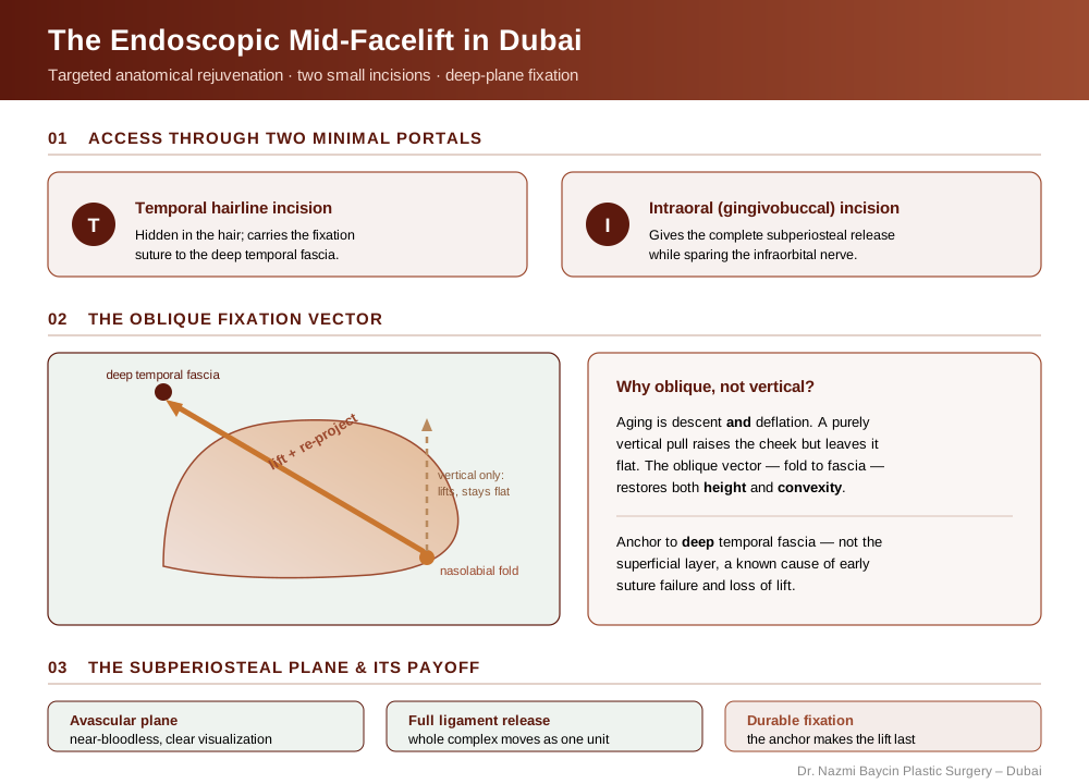
An infographic of the endoscopic mid-facelift in Dubai: the two access portals, the oblique fixation vector that both lifts and re-projects the cheek, and the subperiosteal plane that makes the lift complete and durable — by Dr. Nazmi Baycin, Dubai.
Incision strategy and access: minimal scars, maximal access
The purported benefit of endoscopic surgery is minimal scarring, but poor incision planning compromises the whole operation. My standard approach uses two access points: a temporal incision within the hairline and a lateral gingivobuccal incision. The temporal incision must be placed precisely — too anterior risks the frontal branch of the facial nerve, while too posterior limits instrument angulation for effective mid-face manipulation. The intraoral incision is indispensable in my protocol for a complete subperiosteal release.
The critical consideration is the level of dissection relative to the infraorbital nerve. I use a nerve-sparing technique under direct endoscopic visualization to release the periosteum over the zygoma and maxilla while preserving the nerve’s exit point and its sensory function. This controlled access is what allows major soft-tissue repositioning through truly minimal portals.
The dissection plane: subperiosteal versus supraperiosteal
The choice of surgical plane is the central technical decision in an endoscopic mid-facelift in Dubai. Each option has distinct trade-offs, summarized below.
| Consideration | Subperiosteal plane | Supraperiosteal (deep midfacial) plane |
|---|---|---|
| Best for | Significant anterior projection & vertical elevation | Less severe descent |
| Release | Complete release of all retaining ligaments | More selective malar-pad lift |
| Control of drape | Robust; whole complex moves as one unit | Less powerful overall control |
| Swelling | More, from wider dissection | Potentially less |
| Key caution | Wide release along pyriform aperture & infraorbital rim | Exquisite care to avoid buccal-branch injury |
The subperiosteal plane is my choice for most patients needing significant projection and vertical elevation. Dissecting beneath the periosteum offers a robust, avascular plane and a complete ligament release, so the entire mid-facial envelope mobilizes as one unit. My criticism of many poorly executed endoscopic lifts is an inconsistent, hybrid dissection that fails to fully release the critical ligaments, leading to incomplete correction or rapid relapse. My technique commits fully to a wide, controlled subperiosteal release to guarantee the intended vector of lift.
It is only fair to record that the published long-term evidence points the other way on this specific question. A 2019 retrospective review of 143 patients operated by a single surgeon measured a median 3.4 mm reduction in midfacial height after endoscopic midface lift, with 2.1 mm still sustained between five and fifteen years later. That series used the preperiosteal plane, and its authors concluded that surgery which minimally disrupts the zygomatic and orbicularis retaining ligaments can deliver long-lasting improvement. I continue to favour a complete subperiosteal release in patients needing genuine anterior projection, but the choice of plane is a live question rather than a settled one, and I weigh it case by case.
Fixation biomechanics: ensuring longevity of the repositioned tissues in Dubai
The innovative aspect of the endoscopic approach is not the access, but the method of fixation. Once mobilized, the mid-face complex must be secured under appropriate tension to a stable anatomical point. I exclusively use the deep temporal fascia as my fixation point, for its strength and its posterior-superior vector that best recreates the youthful cheek.
The details are paramount. I use a non-absorbable, braided suture for permanent fixation, since monofilament can cut through delicate tissue under tension. The suture must take a substantial bite of the elevated tissue — malar periosteum and SMAS — and anchor to a robust segment of deep temporal fascia; anchoring to superficial temporal fascia is a recognized error that leads to early suture failure and loss of lift. The vector is not purely vertical, but oblique, running from the nasolabial fold to the lateral canthus, so it elevates and re-projects the cheek at once — correcting both the fold and the flatness.
That fixation, in my experience, is what separates a lasting result from a temporary one. For a fuller view of how I tailor the plan to individual anatomy, see my approach to facelift surgery in Dubai.
Complication avoidance: nerve management and hemostasis
The margin for error in endoscopic surgery is small, and two priorities guide my intraoperative conduct.
- First, facial nerve preservation: the endoscopic corridor places the zygomatic and buccal branches at risk during dissection and fixation, so my protocol relies on constant anatomical visualization, blunt dissectors, judicious bipolar cautery on low settings, and nerve stimulation as a routine check before placing fixation sutures in the lateral mid-face.
- Second, meticulous hemostasis: a bloodless field is a surgical necessity for visualization, not a preference. I use tumescent infiltration with vasoconstrictors and systematically check the internal maxillary artery perforators and the angular vessel before closure, because a postoperative hematoma in this confined space can cause prolonged edema or fibrosis and compromise the final contour.
Synergy with adjacent procedures: the integrated facial framework
An isolated mid-facelift can create disharmony if the periorbital and lower-facial units are unchanged, so my planning always treats the face as an integrated structure. For concomitant brow ptosis, I integrate an endoscopic brow lift in Dubai through the same temporal port, sharing the dissection plane and fixation point for synergistic lifting. For lower-lid aging or lid-cheek junction irregularities, a blepharoplasty in Dubai blends the transition seamlessly.
For patients with cervical laxity, the mid-facelift can be staged or combined with a precision neck lift under a separate plan to manage surgical time and recovery. Because each plan is individual, its details and cost are discussed transparently at the planning consultation rather than quoted from a menu. This architectural philosophy runs through all of my facial plastic surgery work in Dubai.
The endoscopic mid-facelift as a paradigm of modern surgical art
The endoscopic approach to the mid-face is not a shortcut. It is a more demanding, more precise discipline that offers a superior solution for the appropriate candidate in Dubai. It asks of the surgeon a fusion of detailed anatomical knowledge, proficient hand-eye coordination for screen-based dissection, and the artistic judgment to apply the correct vectors of lift. For the informed patient, it represents the evolution of facial rejuvenation — from blunt traction to architectural restoration.
FAQs about the endoscopic mid-facelift in Dubai
-
Who is the ideal candidate for an endoscopic mid-facelift rather than a full facelift?
The ideal candidate is in their late 30s to early 50s with early to moderate mid-facial descent — specifically a flattening of the cheek projection and deepening of the nasolabial folds — but without significant jowling or neck laxity. If the primary aging is concentrated in the mid-face and the lower face and neck remain relatively youthful, an endoscopic mid-facelift addresses the problem precisely without the recovery burden or scar distribution of a full facelift. Patients with more advanced jowling or neck changes are better served by a combined or full facelift approach.
-
How is the endoscopic approach different from a traditional mid-facelift incision?
A traditional mid-facelift uses a longer incision in front of the ear, with direct access to the SMAS layer. The endoscopic approach achieves comparable or superior soft tissue repositioning through two small access points — a temporal hairline incision and an intraoral incision — with a camera-guided subperiosteal dissection rather than direct open access. The trade-off is technical complexity: the surgeon operates in a small field on a two-dimensional screen, requiring specialized instrumentation and a higher level of spatial precision. The benefit is dramatically less visible scarring and typically a faster recovery.
-
What does subperiosteal mean, and why does it matter for longevity?
The periosteum is the dense fibrous membrane that envelops bone. Dissecting beneath it separates the entire soft tissue complex — skin, fat, muscle, and SMAS — from the skeleton as a single unit. This plane is naturally avascular (nearly bloodless) and allows the full release of all retaining ligaments that tether the descended tissue to the bone. When the entire complex is released and repositioned, the lift is more complete and more durable than approaches that work superficial to the periosteum. Endoscopic midface lifting has objective long-term data behind it, with measured improvement in midfacial height still present years after surgery.
-
Why is the fixation vector oblique rather than simply vertical?
A purely vertical lift elevates the cheek but does not address the anterior deflation — the flatness — that contributes equally to mid-facial aging. An oblique vector running from the nasolabial fold to the deep temporal fascia simultaneously elevates the descended tissue and re-projects it forward, recreating both the height and the convexity of the youthful cheek. This is the same principle that distinguishes a deep-plane facelift from a skin-only facelift: the vector must match the direction of aging, not simply oppose gravity.
-
What is the risk to the infraorbital nerve, and how is it managed?
The infraorbital nerve exits the maxilla through the infraorbital foramen — which lies within the subperiosteal dissection zone. If the periosteum is stripped carelessly over this exit point, the nerve can be bruised or stretched, causing temporary numbness across the cheek and upper lip. My protocol involves direct endoscopic visualization of the foramen before and during dissection over that area, using blunt instruments rather than sharp ones in this region, and releasing the periosteum in a controlled circumferential manner around the nerve rather than directly across it. Temporary numbness lasting weeks is possible; permanent nerve injury is exceedingly rare with this level of care.
-
How much downtime should I expect, and when will I see the final result?
Significant swelling and bruising around the cheeks and eyes peaks at three to five days and substantially resolves within two to three weeks. Most patients feel comfortable returning to professional and social settings within ten to fourteen days. The mid-face can appear slightly over-corrected or tight in the first six to eight weeks as residual swelling resolves — this is expected and part of the planned result. Final contour, where all swelling has settled and the repositioned tissue has fully integrated, is visible at three to six months.


GET APPOINTMENT
Get ready to look and feel best… You deserve…
