

Key takeaways: when the scan decides the surgery
- Gynecomastia surgery succeeds or fails on one distinction: gland versus fat.
- Palpation is usually enough — but not always; a muscular or thick chest wall can hide the truth.
- High-resolution ultrasound maps the tissue: gland-to-fat ratio, disk size, fibrosis, and vessels.
- Fat-dominant chests need liposuction; gland-dominant need excision; mixed need both.
- A scan is selective, not routine: it is for the chests palpation reads least reliably.
- For athletic, lean men, imaging enables the millimeter-level precision the result demands.
This is not merely a matter of preference. The imaging literature supports tailoring the operation to the tissue; a 2020 review of male breast imaging identifies pseudogynecomastia as one of the principal differential diagnoses of true gynecomastia, and sets out the imaging appearances that separate them — a distinction that matters clinically, because fatty pseudogynecomastia is usually treated by liposuction alone while glandular tissue requires excision. Ultrasound is how that distinction is made with confidence before a single incision.
The limits of palpation: when feeling is not enough
Many practitioners rely on physical examination alone, and in a slim chest with an obvious disc beneath the areola the examination is usually conclusive on its own. But palpation is inherently subjective, and in some chests it can be genuinely misleading. A muscular chest wall, a thicker subcutaneous layer, or diffuse tissue composition can all disguise what the hands feel. The common errors that follow are predictable: mistaking dense fat for gland, missing deep-seated glandular extensions that reach beyond the areola, and underestimating asymmetry between the two sides.
Each of these translates directly into a surgical misstep — performing liposuction where excision is required, or leaving glandular tissue behind that produces residual puffiness once the swelling settles. When the diagnosis is a guess, the operation is a gamble. That is why, in Dubai, I scan whenever the examination is anything less than conclusive, rather than proceeding on instinct.
In practice that means imaging is selective rather than automatic. I reach for it in the chests where examination is least dependable: a muscular or thick chest wall, a long-standing case where fibrosis may have set in, visible asymmetry between the two sides, a lean athletic patient where the margin for error is smallest, and any revision case where previous surgery has already altered the anatomy.
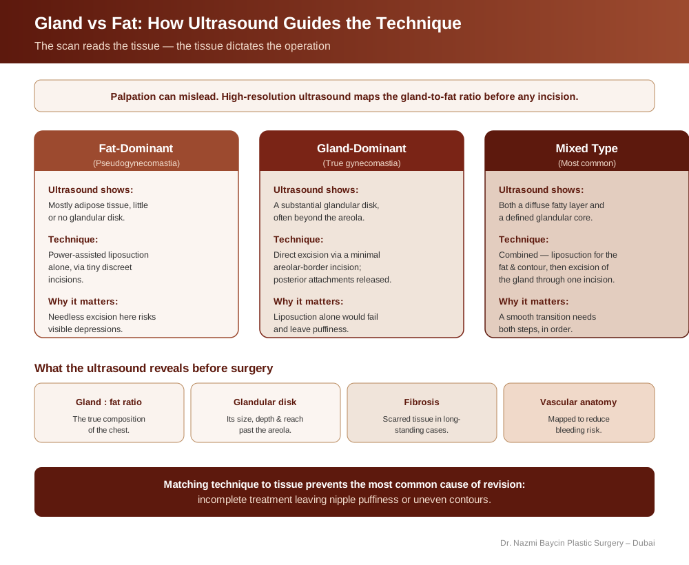
How ultrasound guides gynecomastia technique by tissue type — fat-dominant chests treated with liposuction, gland-dominant with excision, and mixed with a combined approach — after the scan maps the gland-to-fat ratio, glandular disk, fibrosis, and vascular anatomy — by Dr. Nazmi Baycin, Dubai.
The ultrasound advantage: seeing what lies beneath
A high-resolution ultrasound provides a real-time map of the chest wall, showing clearly what palpation cannot reliably discern. It reveals the exact composition and ratio of fatty versus glandular tissue; the precise dimensions, depth, and shape of the glandular disk, which often extends well beyond the border of the areola; the presence of fibrosis in long-standing cases, which calls for a different surgical technique; and the critical vascular anatomy that lets me plan a safer procedure with minimal bleeding risk.
This imaging turns the consultation from a discussion of possibilities into a review of concrete facts. When we can see the exact cause of the contour, we can design the exact solution — the standard I hold for gynecomastia surgery in Dubai, rather than opening the chest and adapting on the spot.
What the scan changes about the plan
The ultrasound findings directly dictate the operative plan. There is no universal gynecomastia operation; there is only the correct procedure for a specific anatomy. The table below summarizes what each imaging pattern means for the decision — the techniques themselves, and how each is performed, I set out in my article on the three types of gynecomastia and the operation each one needs.
| Tissue Pattern | Ultrasound Finding | Surgical Technique | Key Risk if Misjudged |
|---|---|---|---|
| Fat-dominant (pseudogynecomastia) | Mostly adipose, minimal gland | Power-assisted liposuction alone, tiny incisions | Needless excision creates depressions |
| Gland-dominant (true gynecomastia) | Substantial glandular disk | Direct excision via areolar-border incision | Liposuction alone fails, leaves puffiness |
| Mixed type (most common) | Diffuse fat plus a glandular core | Liposuction first, then excision through same incision | Skipping a step leaves contour or lump |
| Long-standing / fibrotic | Fibrosis within the tissue | Modified technique for scarred tissue | Standard approach may under-treat |
| Athletic / lean | Thin fat, gland highly visible | Millimeter-precise, conservative resection | Over-resection causes hollows |
Two of these rows exist only because of the scan. Fibrosis in a long-standing chest and a highly visible gland under a thin fat layer are both findings that change the resection before a blade is picked up, and neither is reliably apparent from the outside. What the anatomy underneath actually consists of — gland, superficial fat, and the deep layer over the muscle — I explain in my article on what gynecomastia is made of.
To understand the full treatment in detail, you can explore gynecomastia surgery in Dubai. Once the tissue is accurately diagnosed, the same principles carry through to building a naturally masculine chest contour.
The athletic and lean patient: a case for precision
For athletic men with low body fat, the margin for error is close to zero. Because there is so little fat to conceal any imperfection, even a small remnant of gland or a slight over-resection of fat can create a visible deformity. Here ultrasound is invaluable. It allows millimeter-level planning, so I can remove just enough tissue to reveal the underlying pectoral anatomy without creating unnatural hollows or asymmetry. This is the precision that delivers the defined, athletic result these patients seek in Dubai — and it is very difficult to achieve reliably when the plan is based on palpation alone.
Beyond technique: the psychological value of an accurate diagnosis
Gynecomastia carries a real psychological burden. Men often describe years of avoiding beaches, pools, and fitted clothing, and many have spent long hours in the gym trying to fix a problem that exercise simply cannot resolve, because glandular tissue does not respond to training. Showing a patient an ultrasound image that clearly demonstrates the glandular tissue is profoundly validating.
For my gynecomastia patients in Dubai, it moves the conversation from subjective worry to objective medical fact, which builds trust and sets realistic expectations about what surgery can and cannot achieve. Understanding exactly what is being treated, and why, changes how a man approaches the whole process.
The first cut is made by the ultrasound probe
Successful gynecomastia correction begins not with a scalpel but with a transducer. In a field where many promise transformation, I believe in definitive diagnosis: by declining to guess, and by looking at the anatomy whenever the examination leaves room for doubt, I make the surgical strategy as individual as the patient. Imaging in those cases is also what guards against the most common reason for revision surgery — incomplete treatment that leaves persistent nipple puffiness or uneven contours.
For any man tired of concealing his chest, the path to a confident, permanent result often starts with this simple, non-invasive scan, and the same diagnostic rigor informs all of my plastic surgery practice in Dubai. Because each anatomy is different, the surgical plan and its cost are discussed transparently at consultation, once the ultrasound findings are reviewed together.
FAQs about gland-vs-fat assessment in gynecomastia
-
What is the difference between gland and fat in gynecomastia?
Gynecomastia refers to enlargement of the actual glandular breast tissue in men, while pseudogynecomastia is chest fullness caused by fat alone. The distinction matters enormously because the two respond to completely different treatments: fatty (pseudogynecomastia) tissue can usually be corrected with liposuction, whereas true glandular tissue generally has to be surgically excised. Glandular tissue also does not shrink with diet or exercise, which is why many men cannot resolve it in the gym. Determining the exact balance of gland and fat is the foundation of planning an effective operation.
-
Why is ultrasound used before gynecomastia surgery?
Ultrasound gives an objective, real-time map of the chest wall that physical examination cannot match. It shows the exact ratio of fat to gland, the size and depth of the glandular disk, whether fibrosis is present in long-standing cases, and the vascular anatomy needed to plan a safer procedure. I do not scan every patient — in a slim chest with an obvious subareolar disc the examination settles it — but where palpation can be misled by a muscular chest or a thick fatty layer, imaging converts the diagnosis from an educated guess into concrete fact. That precision is what allows me to select exactly the right technique rather than adapting mid-surgery.
-
Can’t the surgeon just tell by feeling the chest?
Often, yes — and in a straightforward chest the examination is conclusive. The difficulty is that palpation is inherently subjective, and several factors can obscure what the hands feel — a muscular chest wall, a thicker subcutaneous fat layer, or diffuse tissue composition. This leads to predictable errors: mistaking dense fat for gland, missing deep glandular extensions beyond the areola, or underestimating asymmetry. Any of these can cause a surgical misstep, such as performing liposuction when excision is truly needed. Confirming the plan with imaging in exactly those chests is what avoids these mistakes and the revision surgery that often follows them.
-
How does the tissue type change the surgical technique?
The findings dictate the operation. For a fat-dominant chest with minimal gland, power-assisted liposuction alone through tiny incisions gives an excellent contour, and aggressive excision would only risk depressions. For a gland-dominant chest, liposuction will not work; the glandular plaque must be excised through a minimal areolar-border incision, with its posterior attachments released to prevent recurrence. For the mixed type — the most common — liposuction defines the fatty contour first, then the glandular core is excised through the same incision. One diagnosis, three quite different operations.
-
Why do lean and athletic patients need especially careful assessment?
In athletic men with low body fat, there is very little tissue to hide any imperfection, so the margin for error is almost nil. A small remnant of gland can remain visible, and even slight over-removal of fat can create an obvious hollow or asymmetry. Ultrasound allows millimeter-level planning, so I can remove precisely enough tissue to reveal the underlying pectoral definition without creating unnatural depressions. For these patients in particular, imaging is what makes a crisp, natural, athletic result achievable and repeatable.
-
Does accurate diagnosis reduce the chance of needing revision surgery?
Yes. The most common reason for revision gynecomastia surgery is incomplete treatment — tissue left behind that shows up as persistent nipple puffiness or uneven contours once swelling resolves. This usually traces back to an inaccurate initial diagnosis, such as treating a gland-dominant chest as if it were purely fatty. By mapping the tissue precisely with ultrasound and matching the technique to it, the aim is a single, definitive procedure that addresses everything the first time, reducing the physical, emotional, and financial cost of a second operation.
-
Is the ultrasound assessment uncomfortable or invasive?
Not at all. Diagnostic ultrasound is quick, painless, and completely non-invasive — the same safe technology used in many routine medical assessments. A probe is moved gently across the chest to produce real-time images, with no needles, radiation, or recovery time involved. Beyond its diagnostic value, many men find it reassuring to actually see the tissue on screen, because it explains clearly why they have not been able to resolve the problem on their own and what the surgery will specifically address. It is a simple step that meaningfully improves the reliability of the result.


GET APPOINTMENT
Get ready to look and feel best… You deserve…
